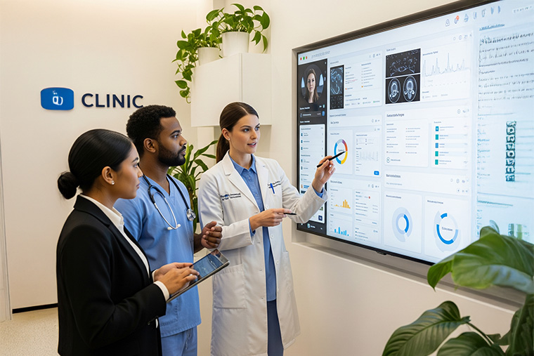
Revolutionizing Healthcare with Medical Image Analysis

Cabot leverages AI-powered tools and deep learning to enhance image analysis, helping you diagnose, treat, and monitor patient conditions with precision.

Transforming Diagnosis with Medical Image Analysis

Medical image analysis allows physicians to spatially investigate a disease site image like CT Scan or Ultra sound, enabling precise intervention for diagnosis and treatment, and to observe particular aspect of patients’ conditions that otherwise would be hard to notice. With the advance of AI, Deep learning and machine learning are used in image analysis to enhance images, detect objects, and classify images.

Types of Medical Images

Tools Available for Image Analysis

sync_alt

AMIDE

AMIDE is a free tool for viewing, analysing, and registering multimodality volumetric medical imaging data sets. AMIDE can also be used to format and export static or moving images. AMIDE can open various types of 2D and 3D imaging data. AMIDE runs on UNIX, Macintosh OS X, and Microsoft Windows platforms, and it is freely available with source code under the terms of the GNU General Public License.

analytics

FSL

FSL is a software library owned by The University of Oxford, that offers many analysis tools for MRI brain imaging data. It is Licenced for non-commercial use for the benefit of educational and research purposes. It includes tools for linear image registration (FLIRT), non-linear image registration (FNIRT), automated tissue classification (FAST) and many others.

trending_up

ImageJ

ImageJ, a Java-based image processing program, offers a broad range of tools for both 2D and 3D images. Beyond standard operations like filtering, edge detection, and resampling, it also includes more advanced image analysis algorithms. ImageJ supports various common 2D image formats and can also open DICOM files used in medical imaging.

admin_panel_settings

ITK-SNAP1

ITK-SNAP is a free, open-source image segmentation tool with a graphical user interface, used for navigating and segmenting 3D medical images. It incorporates automatic segmentation algorithms based on active contours. The software features a dynamic interface for 3D image navigation and offers several manual delineation tools. ITK-SNAP supports a variety of 3D image formats, such as DICOM, NIfTI, and Analyze. ITK-Snap has an intuitive UI and is adept at creating lesion masks on MRI images. ITK-Snap performs semi-automatic segmentation where the structure of interest looks very similar to some of the adjacent structures.

Facing Complex Diagnostic Challenges? Let Cabot Provide the Solution!

Why Choose Cabot for Medical Image Analysis Solutions?


Custom AI Models

Tailored deep learning algorithms for accurate image segmentation, anomaly detection, and classification.


Regulatory Compliance

All solutions are HIPAA-compliant, ensuring the highest standards of patient data security.


Scalable, Real-Time Solutions

Designed to handle high data volumes, our solutions cater to both small clinics and large hospital networks, delivering real-time, data-driven insights for faster and more informed decision-making.

Facing Complex Diagnostic Challenges? Let Cabot Provide the Solution!